Patient Centered Care
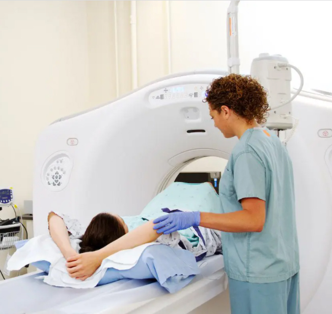
Our Imaging Department is designed to maximize your comfort. Our friendly staff is dedicated to making your experience here as pleasant as possible. For the convenience of our patients and their referring physicians, we provide:
- Easy and flexible scheduling, including some walk-in appointments
- Convenient complimentary valet parking
- Comfortable waiting areas
- Prompt delivery of test results
Faster, Better Communication
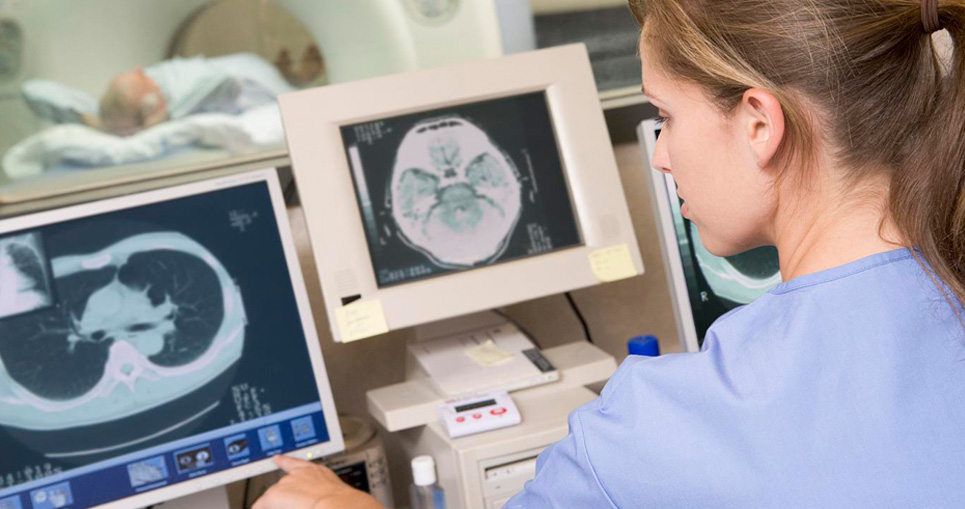
Your doctor has immediate access to the results of your diagnostic procedure — and can view them on a computer screen using secure Internet access from home, the office or the hospital. Our electronic archiving also makes your entire hospital record available to authorized staff at the click of a computer button. If you come back to the hospital with an emergency, your information is instantly available to your doctors and nurses for faster care.
Preparing for Your Imaging Procedure
If you have a scheduled appointment, please discuss the procedure and any special instructions with your doctor before you arrive. If a test is ordered while you are a patient at the hospital, your physician, nurse or technician can explain the procedure and answer any questions you have. These general guidelines will help you prepare for and stay comfortable during your procedure:
- Wear loose-fitting clothes. You may be asked to change into a hospital gown.
- Don’t wear items containing metal. In some cases, you will be asked to remove jewelry, watches, hairpins, eyeglasses or dentures.
- Some tests require you to go without eating or drinking for a specified period before your procedure. Be sure to ask your doctor about this at least 24 hours before your appointment.
- Tell your technologist if there is a chance you may be pregnant or if you are breastfeeding.
- Tell your technologist if you are allergic to iodine or shellfish.
- Some procedures require you to remain still for a period of time. Your technologist will explain what you need to do for a successful procedure.
Advanced Imaging Technologies
North Vista Hospital uses sophisticated imaging technology to diagnose and treat a wide variety of medical conditions.
CT Scanner
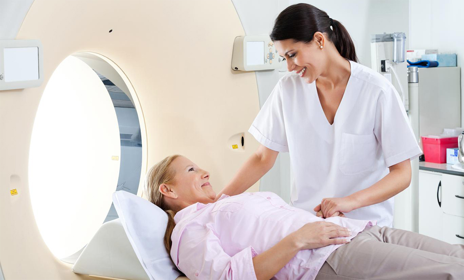
Computer Tomography (CT) scans create images of internal organs, tissues and bones. In just seconds, this advanced technology can make a complete scan of your entire body or produce detailed views of a targeted area, such as the brain or heart. Physicians can view the individual images (called “slices”) for specific detail. Or they can assemble the images to create a three-dimensional view of an organ. The result often is a faster, more precise diagnosis.
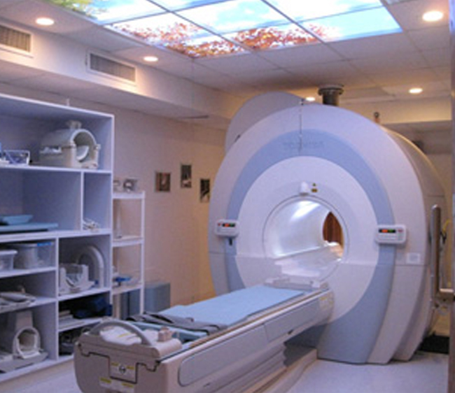
Magnetic Resonance Imaging
MRI technology relies on a combination of radio waves, computer technology and a strong magnetic field to produce high-definition, three-dimensional images of internal organs and structures. Physicians order MRIs to diagnose a wide range of conditions, from cancer, heart and vascular disease and strokes to joint and muscle disorders.

Nuclear Medicine
Nuclear medicine uses tiny, safe levels of radioactive liquid to help physicians diagnose disease. The liquid is formulated so that after you drink it or receive it through an IV, it goes to the part of the body being studied. Then, special cameras scan the affected area. The cameras can detect disease by registering metabolic changes made visible by the liquid. Nuclear medicine procedures are painless and you are exposed to no more radiation than you would receive from a traditional X-ray.

General X-ray
X-rays are the most traditional form of diagnostic imaging. Even with the advent of many newer and more highly advanced technologies, X-rays remain an important part of our Imaging Department and are commonly used to view broken bones and other medical conditions.

Radiographic/Fluoroscopic (R&F) Unit
Radiographic/Fluoroscopic equipment is most commonly used for upper gastrointestinal and barium studies to detect the cause of digestive problems. We also use this technology to detect spinal cord abnormalities, fractures of vertebrae, and certain internal organ functions.
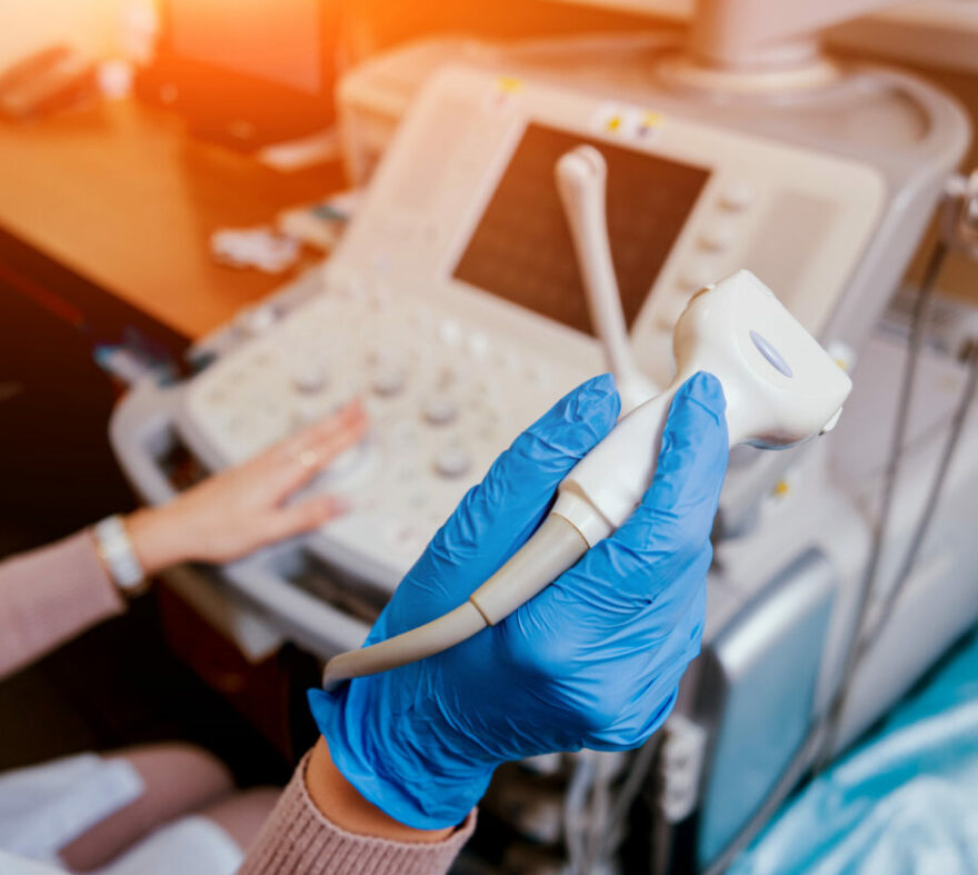
Ultrasound
Ultrasound uses high-frequency sound waves to produce images inside the body that can be viewed on a monitor. It’s a safe and painless way to view the health and development of a baby before birth. Ultrasound can be used to view other areas of the body, including the heart, liver, bladder, kidneys and breasts.
Featured Services
Bariatric & Weight Loss

Emergency Medicine
Wound Care & Hyperbaric Medicine
Mental Health, Adult and Geropsych

Surgical Services


